Biomedical Imaging
Zien is geloven
Geweest
Video's
Podcasts
Artikelen
Over deze serie
Engelstalig symposium
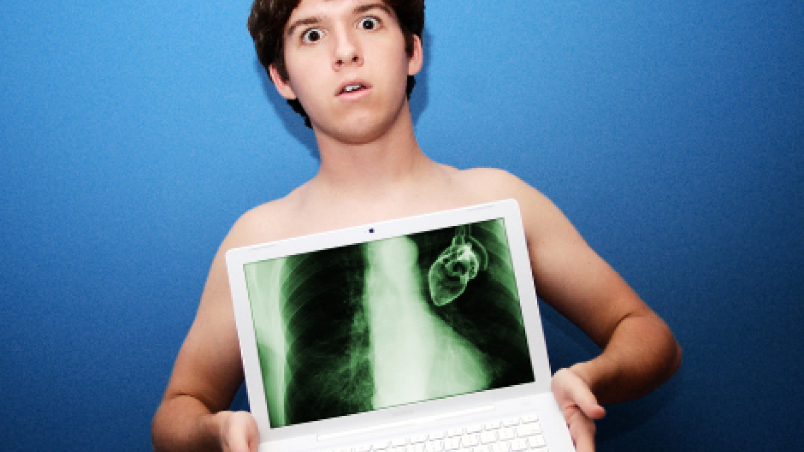
De ontdekking van röntgenstralen in 1895 was revolutionair. Niet langer hoefden artsen te snijden om de mens van binnen te zien. Nu zijn er onder andere CT- en PET-scans, en dé doorbraak van de laatste 20 jaar: de MRI-scan. Met deze technieken kan de wetenschap steeds meer van ons inwendige in beeld brengen.
Maar wat zie je eigenlijk op een hersenscan of een röntgenfoto? Voor onderzoekers en artsen is het interpreteren van beelden een kunst op zich. Het lijken betrouwbare afbeeldingen van wat zich van binnen afspeelt, maar het zijn animaties van hersenactiviteit of andere prikkels. Nemen computers het interpreteren over en kunnen ze dan ook snijden? Hoe geven de beelden inzicht in de vaak voorkomende medische aandoeningen van onze tijd, zoals hersendegeneratie en verschillende kankersoorten zoals borst- en prostaatkanker?
De verwachtingen van beeldvormende technieken zijn hooggespannen. Steeds vaker vragen patiënten om een scan. Welke rol gaat medische beeldvorming spelen bij diagnose, therapie, screening en zelfs tijdens de behandeling? Wat doen we vervolgens met de bergen data die gegenereerd worden uit alle scans en foto's?
Onderzoekers uit verschillende disciplines van de beeldvormende wetenschap gaan tijdens dit symposium in op recente bevindingen omtrent medische scans, diagnose en behandeling. Het symposium wordt georganiseerd in samenwerking met de Graduate School of Life Sciences en geldt voor biomedische masterstudenten als LS-seminar.
15:30 Prof. dr. Max Viergever (UMC Utrecht) - Introduction
15:35 Peter Luijten, PhD (UMC Utrecht) - Ultra High Field MRI
16:00 Prof. Jan Lagendijk, PhD (UMC Utrecht) - MRI-Guided Radiotherapy
16:25 Prof. dr. Paul Suetens (KU Leuven) - Quantitative Image Computing
17:00 Closure
Ultra High Field MRI - Technical challenges, clinical applications
Historically, the magnetic fields strengths used in magnetic resonance imaging (MRI) have shown an almost linear increase over time, for preclinical as well as clinical systems. In the late eighties the first "clinical" 4T systems were introduced. Originally spectroscopy (metabolism) was positioned as the main application for these first generation high field systems, but BOLD based functional brain imaging, looking at the "brain at work", became the most important application in the high field MRI community. Similarly, the recent introduction of even higher field systems (7 and 9.4T), is predominantly driven by the quest for better images for neurocognitive sciences. However, already new, less anticipated clinical applications of ultra-high field systems are being reported. Many of these new applications are related to brain imaging (the detection of new surrogate endpoints for cognition studies, detection of micro infarcts, demyeliniating cortical lesions, intracranial plaque formations). These applications will provide new insight in brain degeneration, one of the major challenges in a society with an increasing elderly population. Also applications for ultra-high resolution outside the brain (breast, prostate) may provide new insights as well, particularly for oncology applications. This will become important for early detection of disease and the control over new therapeutic approaches. Some of these new applications, their potential and limitations, will be discussed as well their future role in tissue characterization for image guided therapies.
MRI guided radiotherapy
MRI is creating a revolution in radiotherapy. Due to innovations in MRI technology like higher field strengths, multi-sense and better gradients, innovative protocols make new contrasts visible and clinically applicable. This multi-parameter imaging of MRI allows tumour characterization and treatment response assessment. The next step is using MRI for guidance of the actual treatment. Unique is the UMC Utrecht development of the MRI linac system, for real-time and on-line guidance of external beam radiotherapy. This MRI linac system has been developed in the UMC Utrecht and is now clinically being introduced in close collaboration with Elekta and Philips. The MR-linac shows the actual soft tissue anatomy during the radiation delivery. The system allows highly precise dose delivery and must be capable to improve local control while minimizing toxicity. The system guides a complete imaging research line on geometrical correct imaging, automatic imaging, real time imaging, automatic segmentation, etc. New clinical procedures are being developed and must be evaluated in clinical studies. MRI guidance is even more important for proton radiotherapy, where the exact depth of the Bragg peak must be controlled real time. We developed the Centre of Image Guided Oncological Interventions (CIGOI). Besides this MRI linac the CIGOI develops MRI guided brachytherapy, MRI guided High Intensity Focused Ultrasound (HIFU) and MRI guided Holmium radio-embolization. Bringing the concept of real time treatment guidance at a new level. Tumour characterization is being developed within the high field MRI group. The presentation during this symposium will give a short introduction to this new treatment concept.
Quantitative Image Computing
The role of imaging in healthcare is continuously increasing. Today, medical imaging plays a role in early patient diagnosis, individualized therapy planning, population screening, therapy outcome prediction and assessment, and translational pre-clinical and clinical research. Recent innovations in medical imaging technology have created a tsunami of imaging data, which is revolutionizing diagnosis, therapy planning and follow-up, as well as clinical, preclinical and biomedical research. Moreover, the rapid adoption of digital image archiving and communication makes that large image databases are readily available for multi-modal, multi-temporal, and multi-subject assessment. A consequence is that accurate quantitative image computing has become indispensable. A prerequisite for quantitative image computing is the availability of suitable models that incorporate prior knowledge about the image content and other patient data, including clinical and genetic information. A powerful strategy is to construct such models from the data itself by learning from a representative training set of image instances. In this seminar several clinical applications of this approach will illustrate the opportunities for population and disease modeling, therapy outcome prediction, evidence-based medicine, and predicting missing or unobserved data.